Social Icons

Pages

Friday 25 December 2009

Summary

The end of the year is usually a period when many people like to summarise their achievements and make future plans. I've been using google analytics for slightly more than 3 months now. It's really great to be able to see the statistics about one's blog.

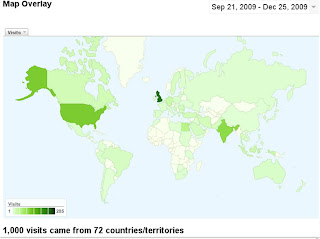
Dental-materials.blogspot.com had exactly 1000 visits between Sept 21 and Dec 25 from 72 countries/territories. The list of top 10 countries with the greatest number of visits includes the USA, India, Egypt, Germany, Indonesia, Canada, Turkey and Iran.


Of 702 absolute uniqe visitors, about one third were returning visitors with more than 300 visits :-) The average time on site was 00:02:26 with an average of 1.90 Pages/Visit.

More than half of all visits came through search engines, Google being the most popular, as expected. About the same number of visits came as direct traffic and from referring sites, such as blogger.com, twitter.com, blogcatalog.com, linkedin.com, facebook.com etc.


Interestingly, this blog appeared in search engines not just when 'dental materials' were used as keywords. In fact, the visitors of this blog entered more than 400 keywords, such as 'dental materials journal', 'mta dental material', 'filtek silorane', 'mta in endodontics', 'free dental material journal', 'books of dental materials' etc.

So, it's been an exciting period for dental-materials.blospot.com and I hope this blog will be even better in the future. I'd like to thank all the visitors and wish Merry Christmas and a very happy and prosperous New Year.

Saturday 12 December 2009

News from jobs.co.uk

PhD Scholarship

Manufacturing And Characterisation Of Titanium Based Dental Implant

University of Sheffield - Department of Mechanical Engineering

Application deadline: 07 January 2010.

Thursday 10 December 2009

Call for abstracts: 88th General Session & Exhibition of the IADR

88th General Session & Exhibition of the IADR
Barcelona, Spain, July 14-17, 2010.
www.iadr.org

Abstracts can be submitted online HERE. The deadline for abstract submission is February 5, 2010. As on previous IADR conferences, one person may present only one abstract but can be co-author on an unlimited number of abstracts.

More than 4500 delegates are expected and more than 3000 abstracts submitted. There will be oral and poster discussions as well as traditional poster sessions. All delegates will receive continuing education credits according to the ADA CERP.

The new way of abstract presentation is through poster discussion sessions designed for up to 7 individual presentations. Abstracts in these sessions will be presented as posters with up to 3 PowerPoint slides in addition. Each presentation will last for 5 minutes followed by 2 minutes of discussion.

Posters will be displayed vertically, rather than horizontally. Poster dimensions will be 0.93 m (width) x 2.22 m (height).

There will be more than 20 subject areas including dental materials, cariology, dental anesthesiology, implantology, microbiology, mineralized tissue, neuroscience/TMJ, oral&maxillofacial surgery, periodontology, prosthodontics, pharmacology, pulp biology etc.

More information and detailed instructions can be found HERE.

Monday 30 November 2009

Poll: What type of adhesive system do you use in your practice?

Please, select one or more answers from the poll in the sidebar.

Adhesive systems are nowadays classified according to the number of clinical application steps and adhesion strategy. This is one of the most widely accepted classifications in both clinical practice and scientific literature.


(a) Three-step etch-and-rinse systems: acid etching with phosphoric acid followed by the application of primer and bond from separate bottles;
(b) Two-step
etch-and-rinse systems: acid etching with phosphoric acid followed by the application of primer and bond which are in one bottle;
(c) Two-step self-etch systems: self-etching primer and bond are in separate bottles;
(d) One-step self-etch systems (also known as "all-in-one" systems): all components are in one bottle

Feel free to leave a comment about your experience with any particular adhesive system.


In clinical practice, I'm using a two-step etch-and-rinse system. In PhD research, I've studied the degree of conversion and elution of unreacted monomers from a wide range of adhesive systems. Some of these results have been published in scientific dental journals and some are in press and will be published soon.

Though one-step (all-in-one) self-etch systems are currently marketed, results from many in vivo and in vitro studies suggest that these systems often have inferior properties compared to two-step self-etch and etch-and-rinse systems


Tuesday 24 November 2009

Textbook of Restorative Dentistry

It is my pleasure and honour to announce that the Textbook of Restorative Dentistry (original title: Osnovi restaurativne stomatologije) has been published by my professors at Belgrade University School of Dentistry Dept. of Restorative Dentistry and Endodontics: Slavoljub Zivkovic, Mirjana Vujaskovic, Katarina Pap, Djurica Grga, Aleksandra Lukic and Nevenka Teodorovic.

This is the official textbook for undergradute students but may be very helpful to dental practitioners who have some questions regarding restorative procedure.

The book is divided in four parts. Part I presents the biology of pulp-dentine complex, its ultrastructure, but also important physiological and biochemical aspects. This chapter also explains the basics of caries etiology, pathogenesis and diagnostics.

Part II details the equipment and instruments for restorative treatment as well as techniques for caries removal.

Part III explains cavity preparation procedures according to traditional Black's principles and the adhesive cavity preparation for resin-based materials. Cavity preparation for indirect restorations is also explained. Some basic gnatological requirements in restorative treatment are mentioned in Part III.

Part IV addresses all dental materials in restorative dentistry. It contains basic clinical characteristics of materials for temporary fillings, for pulp cappings, amalgam, glass-ionomer cements, resin-based composite materials and adhesive systems as well as materials for indirect restorations.

Thursday 19 November 2009

News from jobs.ac.uk

Research Fellow in Tooth and Peridontal Tissue/Biomechanics/Biomedical Engineering
(Click for more information)

Faculty of Engineering
University of Leeds - School of Mechanical Engineering

Salary: £29,704 to £35,469. It is likely that an appointment will be made at or below £31,513 p.a.

Application deadline: 8 December 2009

Monday 16 November 2009

Wiley-Blackwell books: Dental Materials at a Glance [20% off]

A couple of days ago, I got an email from Wiley-Blackwell offering certain dental books at discount, 20% off the original price.

Among the books at discount is "Dental Materials at a Glance" by J. Anthony von Fraunhofer, Professor Emeritus at the Baltimore College of Dental Surgery Dental School, University of Maryland and formerly was Professor and Director of Biomaterials Science in the Department of Oral and Maxillofacial Surgery.

I'm not quite sure why I got this offer, either because I subscribed to the newsletter from Wiley-Blackwell or because I already purchased at least one book. Anyway, I'm considering to buy Dental Materials at a Glance since it's now only $27.19. Having bought another book from this "At a Glance" series (Medical Statistics at a Glance), I assume this book about dental materials is written in a similar succinct and readable way with many useful illustrations.

It contains 28 chapters on 72 pages, i.e. 2-3 pages per chapter, which seems to be the pattern for all books from this series. According to the table of contents, the first part looks like a glossary of scientific terms relevant to any dental materials scientist. The second part seems to be a short overview of important properties of contemporary dental materials. There's also a free downloadable chapter HERE.

Thursday 12 November 2009

Nano-filled resin-modified glass-ionomer cement: "nano-ionomer" Ketac N100

In addition to conventional and resin-modified glass-ionomer cements, a nano-filled resin-modified glass ionomer cement, or “nano-ionomer”, was developed by 3M ESPE a couple of years ago – Ketac N100.

It is stated by the manufacturer that indications for the use of this nano-ionomer include primary teeth restorations, small Class I, Class III and IV, temporary restorations, filling defects and undercuts, “sandwich” technique with resin-based composites, core build-ups with min 50% of the remaining tooth for support.

The nano-ionomer is based on the acrylic and itaconic acid copolymers necessary for the glass-ionomer reaction with fluoroaluminosilicate (FAS) glass and water. The nano-ionomer also contains a blend of resin monomers, BisGMA, TEGDMA, PEGDMA and HEMA which polymerize via the free radical addition upon curing and it is stated that the primary curing mechanism is by light activation. The originality of this glass-ionomer cement is the inclusion of nano-fillers which constitute up to two thirds of the filler content (circa 69 wt%).

Other advantages stated by the manufacturer are a simplified procedure which requires the priming but not the separate conditioning step and a precise dispensing and mixing “clicker” mechanism.

In spite of its uniqueness amongst other dental formulations, the nano-ionomer has not been investigated to a greater extent in the scientific dental literature. Medline search using the keyword “Ketac N100” resulted in only 4 papers in international peer-reviewed journals. Another paper was found using the keyword “nano-ionomer”. It is my pleasure to mention that the first of these 5 papers was done in Serbia by my colleagues from the Paediatric Dept of the School of Dentistry, Belgrade and the Dept. of Dentistry School of Medicine, Novi Sad.

It has been reported that fluoride concentration on material surface is similar for Ketac N100 and other glass-ionomer cements from the Fuji “family” but Ketac N100 showed less porosities and surface cracks than Fuji materials (Markovic et al 2008).

A study on bonding orthodontic brackets showed significantly lower shear bond strength for Ketac N100 compared to a conventional light-cure orthodontic bonding adhesive (Transbond XT). However, it has been suggested that this nano-ionomer may be used for bonding orthodontic brackets since the obtained shear bond strength is within clinically acceptable range (Uysal et al. 2009).

Another study using the shear bond strength as an adhesion parameter showed that Er:YAG laser dentine pre-treatment results in lower bond strength values compared to acid etching or a combined acid-etching and laser pre-treatment (Korkmaz et al. 2009).

A study on microleakage around Class V cavities showed that Er:YAG preparation results in greater microleakage than a conventional cavity preparation with a bur when a nano-ionomer (Ketac N100) and a nano-composite (Filtek Supreme XT) were used as restorative materials (Ozel et al. 2009).

In a study by Leuven BIOMAT Research Cluster it has been concluded that Ketac N100 “bonded as effectively to enamel and dentin as a conventional glass-ionomer (Fuji IX GP), but bonded less effectively than a conventional resin-modified glass-ionomer (Fuji II LC). Its bonding mechanism should be attributed to micro-mechanical interlocking provided by the surface roughness, most likely combined with chemical interaction through its acrylic/itaconic acid copolymers” (Coutinho et al. 2009).

More research is needed to investigate other mechanical properties of the nano-ionomer, its biochemical stability in the oral environment, fluoride release etc. Ultimately, well-designed randomized clinical trials will reveal the longevity and anti-cariogenic effect of this material in clinical conditions.

References:
  • Markovic DLj, Petrovic BB, Peric TO. Fluoride content and recharge ability of five glassionomer dental materials. BMC Oral Health 2008; 28:8-21.
  • Uysal T, Yagci A, Uysal B, Akdogan G. Are nano-composites and nano-ionomers suitable for orthodontic bracket bonding? Eur J Orthod 2009; Apr 28 [epub ahead of print]
  • Korkmaz Y, Ozel E, Attar N, Ozge Bicer C. Influence of different conditioning methods on the shear bond strength of novel light-curing nano-ionomer restorative to enamel and dentin. Laser Med Sci 2009; Aug 18 [epub ahead of print]
  • Ozel E, Korkmaz Y, Attar N, Bicer CO, Firatli E. Leakage pathway of different nano-restorative materials in class V cavities prepared by Er:YAG laser and bur preparation. Photomed Laser Surg 2009; 27:783-789
  • Coutinho E, Cardoso MV, De Munck J, Neves AA, Van Landuyt KL, Poitevin A, Peumans M, Lambrechts P, Van Meerbeek B. Bonding effectiveness and interfacial characterization of a nano-filled resin-modified glass-ionomer. Dent Mater 2009; 25:1347-1357.
Sunday 8 November 2009

Mineral trioxide aggregate (MTA) in endodontics

Mineral trioxide aggregate (MTA) is a mixture of a refined Portland cement and bismuth oxide, and also contains trace amounts of SiO2, CaO, MgO, K2SO4, and Na2SO4. MTA was first described for endodontic applications in the scientific literature in 1993. Nowadays, there are two forms of MTA on the market, the traditional gray MTA (GMTA) and white MTA (WMTA), which was introduced in 2002. WMTA has less Al2O3, MgO, and FeO and, also, smaller particles than GMTA.

MTA is prepared by mixing the powder with sterile water in a 3:1 powder/liquid ratio. This results in the formation of a colloidal gel that solidifies to a hard structure in approximately 3–4h. It is believed that moisture from the surrounding tissues favours the setting reaction.

Similar or less microleakage has been reported for MTA compared to traditional endodontic sealing materials [gutta-percha and pastes] when used as an apical restoration, furcation repair, and in the treatment of immature apices. 3mm of MTA is recommended as the minimal amount against microleakage and 5mm in the treatment of immature apices. In vitro and in vivo studies support the biocompatibility of freshly mixed and set MTA when compared to other dental materials

Clinical applications of MTA include:
pulp capping,
pulpotomy dressing,
root-end filling,
root repair [resorption and perforations] and
apexification.

Clinical prospective studies suggest that both GMTA and WMTA have similar results as traditional calcium hydroxide in non-carious mechanical pulp exposures in teeth with normal pulp tissue. However, further clinical studies are needed, particularly involving pulp exposures in carious teeth.

Clinical prospective studies using MTA as pulpotomy dressings for primary and permanent teeth reported similar or better results for MTA materials compared to formocresol or calcium hydroxide in the formation of dentine bridges and continued root development. Histological analysis has suggested a more homogenous and continuous dentine bridge formation by MTA than calcium hydroxide at both 4 and 8 weeks after treatment and less inflammation associated with MTA than calcium hydroxide.

There are several case reports in which MTA has been successfully used to repair horizontal root fractures, root resorption, internal resorption, furcation perforations and apexification and/or apexogenesis which was confirmed clinically and radiographically.

Overall results on the use of MTA in endodontics are favourable, but more well-designed and controlled clinical longitudinal studies are needed to allow systematic review and confirmation of all suggested clinical indications of MTA.

You may be interested in a list of free full text scientific articles published in international peer-reviewed journals.

Tuesday 3 November 2009

Biocompatiblity of resin-based materials: Bisphenol-A

Bisphenol-A is an organic compound with two phenol functional groups, commonly used in the production of many plastic products. Studies have shown that bisphenol-A may have adverse health effects such as hormonal, developmental, neurological as well as carcinogenic.

In spite of this, bisphenol-A may be part of dental resin-based materials as a direct ingredient, as a by-product of bio-degradation of other ingredients or as a trace material left-over from the manufacture of other ingredients. Though bisphenol-A is rarely used as a direct ingredient in dental composites, adhesives or sealants, it is a starting ingredient in the manufacture of the most commonly used resin - BisGMA. Also, some other, less frequently used resins, such as Bis-DMA, are also produced using bisphenol-A.

Concerns about bisphenol-A have been raised since studies have shown that various components of dental composites, adhesives and sealants may leach out and have potential local or systemic adverse effects. An in vivo study using an ELISA system showed that up to 100 ng/ml of bisphenol-A may be released into saliva from resin-based composites immediately after placement (Sasaki et al. 2005). Also, a more recent study showed that BisGMA, but also BisGA and BisDMA, all bisphenol-A - based monomers, may leach out from in vivo placed composite restorations (Uzunova et al. 2008). On the other hand, no traces of bisphenol-A were found from chemically and light-cured orthodontic adhesives after in vitro accelerated aging and elution (Eliades et al. 2007).

Over the past few years, both the FDA and ADA have supported research on biocompatibility and safety of resin-based materials containing bisphenol-A. Both organizations issued statements in 2007 (FDA) and 2008 (ADA) that the low-level of BPA exposure that may result from dental materials poses no known health threat. However, further research on this subject is encouraged by the ADA.

References:
  • Sasaki N, Okuda K, Kato T. Salivary bisphenol-A levels detected by ELISA after restoration with composite resin. J Mater Sci: Mater in Med 2005;16:297-300.
  • Uzunova Y, Lukanov L, Filipov I, Vladimirov S. High-performance liquid chromatographic determination of unreacted monomers and other residues contained in dental composites. J Biochem Biophys Methods 2008;70:883-888.
  • Eliades T, Hiskia A, Eliades G, Athanasiou AE. Assessment of bisphenol-A release from orthodontic adhesives. Am J Orthod Dentofacial Orthol 2007;131:72-75.

Monday 26 October 2009

Pre-fabricated, direct, single-visit, ceramic inserts

Ceramic inserts have been designed as single-visit systems and an alternative to conventional ceramic restorations produced in two appointments by means of indirect technique. Luting ceramic inserts with a small amount of composite resin is expected to reduce the amount of polymerisation shrinkage by reducing the bulk of resin composite needed to restore the tooth. Another advantage is that the occlusal contacts can be placed on the ceramic surface, rather than on the composite, for longer-term stability.

One of the most studied systems is Cerana, which utilises pre-etched and silanated leucite inlays with matched diamond burs. After caries removal and the preparation of a usual adhesive-type preparation for bonded restorations (Figure 1), the cavity is refined using one of three conical burs (Figure 2). Enamel and dentine are etched if etch-and-rinse adhesive is used or self-etch systems are applied and cured. Composite is then applied to the cavity, filling it to or just above the enamel-dentin junction (Figure 3). A thin coat of composite can be applied to the ceramic insert which is then pressed into the cavity. Excess resin composite is removed and the restoration is cured for 20 s or 40 s depending on the light-curing unit (Figure 4). The occlusal contour of the inlay is shaped to match the surrounding enamel and the occlusion adjusted (Figure 5). The restoration is cured for a further 20 s or 40s and polished.

(Figures from manufacturer's recommendations for use. Nordiska Dental AB, Sweden)
A 3-year prospective clinical trial has shown that “The results indicate that Cerana is an alternative to composite resin restorations in Class I situations, but should be avoided in connection with Class II tunnel preparations.” (Odman 2002)

Another 8-year prospective clinical trial has shown that “Cerana is acceptable in terms of aesthetics, patient acceptance, occlusal wear and ease of use and is a good alternative for a single-visit, tooth coloured restoration in suitable cavity shapes.” (Millar & Robinson 2006)

In an in vitro study Cerana inserts luted with flowable composite in Class V cavities showed significantly less microleakage than those cemented with the high-viscous material only at the gingival margins. Microleakage was reduced around inserts compared to the bulk filling with flowable composites but no difference was observed between inserts and bulk filling with high-viscous composite material (Salim et al. 2005).

It was also shown that in vitro thermocycling 4000 times between 5 and 55 degree C does not increase microleakage around Cerana inserts (Santini et al. 2006). After thermocycling, Cerana inserts showed siginificantly less microleakage along both occlusal and gingival margins compared to Beta Quartz glass-ceramic inserts and Tetric Ceram resin-based composite. Both findings were attributed to the coefficient of thermal expansion of Cerana inserts which approximates that of enamel (Tan & Santini 2005; Santini et al. 2006).

References:
  1. Odman P. A 3-year clinical evaluation of Cerana prefabricated ceramic inlays. Int J Prosthodont 2002; 15: 79-82.
  2. Millar BJ, Robinson PB. Eight year results with direct ceramic restorations (Cerana). Br Dent J 2006; 201:515-520.
  3. Salim S, Santini A, Safar KN. Microleakage around glass-ceramic insert restorations luted with a high-viscous or flowable composite. J Esthet Restor Dent 2005;17: 30-38.
  4. Santini A, Ivanovic V, Tan CL, Ibbetson R. Effect of prolonged thermal cycling on microleakage around Class V cavities restored with glass-ceramic inserts with different coefficients of thermal expansion: an in vitro study. Prim Dent Care. 2006 Oct;13(4):147-53.
  5. Tan CL, Santini A. Marginal microleakage around class V cavities restored with glass ceramic inserts of different coefficients of thermal expansion. J Clin Dent. 2005;16(1):26-31.

Saturday 24 October 2009

News from jobs.ac.uk

Research Fellow in Biomaterials for Skeletal Regeneration
(Click for more information)

Leeds Dental Institute
University of Leeds - Faculty of Medicine and Health

Salary: £29,704 to £35,469 p.a. It is likely that an appointment will be made at or below GBP 31,513 p.a. since there are funding limitations which dictate the level at which the appointment can start.

Application deadline: 3 November 2009

Thursday 22 October 2009

Essential toolkit for a dental materials scientist: Search engine (Part II)

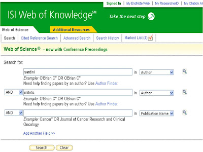
ISI Web of Knowledge (Web of Science)

ISI Web of Knowledge (also known as Web of Science) is a research platform by Thomson Reuters that comprises seven databases containing information gathered from thousands of scholarly journals, books, book series, reports and conferences. It contains three multidisciplinary indexes to the journal literature of the sciences, social sciences and art and humanities; two conference proceedings citation indexes for sciences, social sciences and humanities and two chemistry databases (Index Chemicus and Current Chemical Reactions).

Search panel is very intuitive and user-friendly and allows search by various categories, similar to Medline, such as topic, title, author, title, journal, year of publication, address, conference, language, document type, funding agency and grant number.


A search can be saved under Marked List and forwarded to an email address as plain text or html.

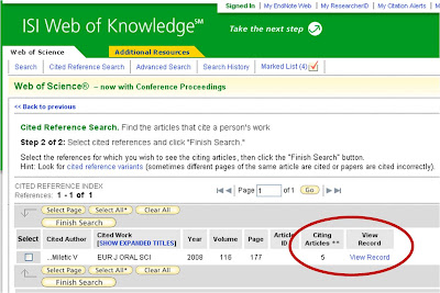
ISI web of Knowledge offers an option to create citation reports which include the total and average number of times certain items (e.g. journal articles) have been cited and also the number of citations per year. Furthermore, ISI Web of Knowledge displays a list of articles in which an article of interest has been cited.


Cited Reference Search is an option to search for articles that cite a certain person’s work. A search can be done using a person’s name, the journal where an article is published and/or the year of publication.


Another useful tool is that EndNote Web version is integrated with ISI Web of Knowledge. This allows a search to be saved as an Endnote bibliography and directly cited while typing. A search can also be exported to other types of Reference software if you have it installed on your computer (EndNote, Reference Manager or ProCite). I will address EndNote as part of the essential toolkit of a dental materials scientist in a separate blog post.

Saturday 17 October 2009

Essential toolkit for a dental materials scientist: Search engine (Part I)

MEDLINE

For many clinical researchers, MEDLINE is probably the starting point for any article search. MEDLINE comprises over 5000 journals published worldwide and is the largest part of the PubMed database, a service of the U.S. National Library of Medicine. PubMed also contains other life science journals.

As a result of U.S. National Institutes of Health Public Access policy aimed at increasing free access to articles, Pubmed Central (PMC) has been created as a free digital archive of biomedical and life sciences journal literature. It contains journals which submit articles regularly but also articles published by NIH-funded researchers in journals currently not on the PMC list. Full text is available in either HTML or PDF format.

A particularly useful tool available at PubMed is “MyNCBI” which allows searches to be saved and filtering options and automatic searches set up. It is located in the top right corner of the PubMed homepage and requires registration (free). MyNCBI offers various features but among the most useful are automatic searches and collections.


Automatic search: Once you enter keywords and search results are generated, you should save the search by clicking the “Save Search” option next to the search box. The search is saved to MyNCBI. Then, you can enable automated search in MyNCBI and the results will be emailed to you daily or once a week or month, according to the settings. The same keywords from the initial search will be used every time in the automated search.

Collection: Once you enter keywords and search results are generated, you should save the search by clicking the “Send to” option and selecting “Collections” from a drop down menu. A collection can be made public by selecting the appropriate option in MyNCBI, in which case a direct URL or HTML for web pages and blogs are generated.

You can access MyNCBI through PubMed homepage, but if no PubMed search is intended, then you can use a direct link to MyNCBI


Friday 16 October 2009

News from jobs.ac.uk

Lecturer in Restorative Dentistry
(Click on the link for more information)

University of Leeds - Leeds Dental Institute
Salary on the Clinical Lecturer scale (£30,685 - £57,084 pa)

Application deadline: 24 October 2009

Monday 12 October 2009

Resin-based materials: Degree of conversion

Resin-based materials, such as resin-based composites, adhesives, pit & fissure sealants and resin cements, undergo monomer to polymer conversion during both light-activated or chemically-activated polymerisation. Whilst conversion is an inherent property of resin-based materials, the degree of conversion (DC) depends on material chemical composition and curing conditions. The DC affects mechanical properties of resin-based materials, such as wear, fracture toughness, hardness, flexural modulus and fatigue. Less than optimal conversion may compromise mechanical properties but also result in leaching of monomers from restorations.

Traditionally, manufacturers' technical and scientific data did not incorporate results of internal or external tests for the DC. Even most recent materials often lack these data but there seems to be a growing understanding of the importance of this property.

The DC of resin-based materials in dental studies has been determined using various methods for more than two decades, but recently, the most widely accepted and used methods are infrared and Raman spectroscopy. These are based on measuring the changes in either the absorbance or scattering effect of those molecular groups which take part in polymerisation of resin-based materials. The DC is determined as the ratio of absorbance or scattering of these groups and a certain internal standard in uncured and cured material. An internal standard is another molecular group which does not take part in polymerisation and, thus, its infrared absorbance or Raman scattering remains constant before and after polymerisation.

It is very important to point out that the DC indicates the number of unreacted methacrylate or other polymerisable groups and not the amount of unreacted monomers in the polymer. The DC of e.g. 70% indicates that there is 30% of uncreacted groups and not 30% of free, unreacted monomers trapped within the polymer network that could theoretically leach out. Cross-linking monomers in resin-based materials most often contain more than one polymerisable group which means that cross-linking may occur via some but not all such groups. Furthermore, unreacted polymerisable groups always exist at the ends of polymer chains. There have been some estimates that in resin-based composites with the DC of around 70%, the amount of unreacted monomers is actually less than 10%. This depends on material chemical composition and may vary significantly in different resin-based materials.

Thursday 8 October 2009

Dental Research in the UK: Funding

In 2006, the British Society for Dental Research (BSDR) commissioned a position paper on oral and dental research within the United Kingdom which would serve as a foundation and a framework for a national plan for oral and dental research. This paper was written by Iain Chapple, Paula Farthing, David Williams and Michael Curtis in 2006 and updated in December 2007.

According to this strategic review, the UK dental school research income in the period 2000-2004 was less than 2% of medical schools research income. Knowing that the NHS spend on dental care was about 5% of the total NHS healthcare spend, this indicates that dental research was under funded compared to medical research.

The majority of dental research funding was provided by UK government, industry and charities over this five-year period. Funding from research councils, EU and other sources constituted up to one third of the overall funding per year. It is interesting that most of the charitable funding came from the Wellcome Trust since there is no national charitable source dedicated to oral research.

The best financed research in UK dental schools has been basic with more than two thirds of awarded grants. Less than one third of grants was awarded for clinical research in dental schools. These data are based on grants awarded between 2000 and 2005 by the Medical Research Council (cca. £6m), Wellcome Trust (£8.68m), Biotechnology and Biological Sciences Research Council (£cca. 1.2 m) and Engineering and Physical Sciences Research Council (£520k). The percentage of dental materials research funding has not been reported in this position paper.

Some of the identified reasons for under funding dental research in UK dental schools include the lack of representation on review panels of research councils and major charities, the absence of a national charitable source for funding oral research and underscoring grant proposals by internal panels in spite of high scores by external expert reviewers.

Four priority areas have been proposed in this position paper:

  • Establishing a dedicated Oral and Dental Research Charity
  • Better representation for oral and dental research on review panels
  • Developing critical mass through nationally-coordinated research consortia and
  • Encouraging inter-school collaboration.

Monday 5 October 2009

Light curing of resin-based composites and adhesive systems

Light cured resin-based materials are predominantly used in current dental practice. Light curing protocols have changed over time following changes particularly in light-curing units (LCUs) since the photoinitiator system in these materials has remained virtually unchanged. Though there are attempts to modify the photoinitiator system, the most frequently used one is based on camphorquinone and a tertiary amine.

On the other hand, the LCU technology has been developing in several directions. LCUs comprise four different types of light sources: halogen, light-emitting diode (LED), plasma arc and laser. Halogen and LED LCUs are most often used in dental practice and studied in the dental literature. Light intensity and curing time have been identified as important parameters in monomer conversion which affect mechanical characteristics of the resultant polymer and subsequently its clinical performance. As light intensity has increased from about 500 mW/cm2 which is characteristic of the so-called ‘conventional’ LCUs to more than 700 mW/cm2 in the so-called ‘high-power’ LCUs, most manufacturers recommend shorter curing time. Consensus opinion in the current dental literature is that light energy density (light intensity multiplied by curing time) is a more important determinant of the degree of conversion of resin-based composites (RBCs) and adhesives than light intensity. It is currently recommended to cure adhesive systems for 20 s with LCUs operating at intensities of about 500 mW/cm2 and 10 s with LCUs operating at intensities of more than 700 mW/cm2. For RBCs, the recommended curing time is 40 s with the former LCUs and 20 s with the latter ones. The recommended thickness for each layer of RBCs in the incremental technique is still 2 mm.
Though many LCUs possess additional curing modes, such as soft-start or pulse in order to reduce polymerisation shrinkage of RBCs, there is no scientific evidence that these modes affect the long-term clinical performance of resin-based restorations.

It has been shown that maximum absorption range of camphorquinone is about 468 nm and therefore most LCUs, especially LED and plasma arc, have a very narrow emission range. However, the absorption range of co-initiators may be outside the emission range of such LCUs, thus, leading to insufficient conversion. Most recently, the so-called ‘poly-wave’ LCUs have been introduced on the market in an attempt to cover the absorption range of the entire photoinitiator system and produce maximum conversion for a given material. Future studies will show whether this new approach ensures such monomer to polymer conversion which would lead to better mechanical properties of RBCs and adhesives.

Studies have shown that increased curing distances lead to lower degree of conversion and it has recently been suggested that 6 mm may be a cut-off distance. However, it should be noted that various LCUs and materials may exhibit differences in curing efficiency at various distances. Therefore, as a general rule, the LCU tip should be placed as close as possible to the surface of RBCs and adhesives.

The superficial layer of RBCs and adhesives is insufficiently cured due to oxygen inhibition. It is removed by polishing RBCs but in adhesives, this layer serves as an intermediate zone enabling the formation of the RBC-adhesive bond. It is, therefore, important to use RBCs and adhesives with compatible chemical composition in order to achieve optimal RBC-adhesive bond by interaction of compatible monomers from both materials.

Saturday 3 October 2009

News from jobs.ac.uk

Postdoctoral Research Associate

Tissue engineering scaffolds for bone tissue engineering
King's College London - Department of Dental Biomaterials Science
Salary: £30,000 to £49,999

Application deadline: October 26, 2009.

Friday 2 October 2009

Filtek Silorane by 3M ESPE

In 2007, 3M ESPE launched a new resin-based composite, Filtek Silorane (FS), and its adhesive system. Both the composite and the adhesive system contain a unique resin monomer based on the combination of siloxanes and oxiranes so it is apparent where the term "silorane" comes from. The polymerisation of FS differs from methacrylate-based composites and adhesives and is claimed to result in reduced polymerisation shrinkage. The cationic polymerisation of FS occurs via the ring opening of the C-O-C epoxide group which ends up in less reduction in molecule distances compared to the free radical polymerisation of methacrylate-based composites. In the latter, monomer interaction via methacrylate C=C groups results in the greater reduction of inter-molecular distances and subsequently greater polymerisation shrinkage.

Most recent studies have shown reduced shrinkage and shrinkage stress and strain for FS compared to methacrylate-based composites. Microleakage and nanoleakage were also reported for FS. Ongoing studies will reveal other properties of FS that may affect its clinical performance.

The dedicated adhesive system is designed to bridge the gap between hydrophilic dentine and hydrophobic FS composite. It contains the Primer and the Bond in separate bottles which are cured as separate layers, unlike any other two-step self-etch adhesive system, where primer and bond are mixed before curing. In Filtek Silorane adhesive system, these layers are not visible on SEM but can be detected using micro-Raman spectroscopy (Santini & Miletic, 2008) At the BSDR symposium on Dental materials it was reported, that after 6 months of storage, the type of failure for FS changes from the adhesive to cohesive as the fracture occurs within the adhesive system. The intermediate zone between FS Primer and Bond of about 1 micron may be the weak link in the failure mechanism and certainly needs further investigation.

Monday 28 September 2009

Recommendations for conducting controlled clinical studies of dental restorative materials

Inspired by the recent debate, I did a literature search on clinical trials in various dental disciplines. As expected, there are loads of such studies on dental materials and clinical procedures, so the argument that something can't be tested is invalid. Everything can and must be tested using scientifically structured protocols before certain claims are made.

Two years ago, a group of scientists associated with the FDI Science Committee published recommendations for conducting clinical trials on dental materials. These recommendations are related to study design and evaluation criteria.

The following is the abstract of this paper and the full text can be obtained from J Adhes Dent or the first author, Dr Reinhardt Hickel of the University of Munich, Germany hickel@dent.med.uni-muenchen.de

Recommendations for conducting controlled clinical studies of dental restorative materials. Science Committee Project 2/98--FDI World Dental Federation study design (Part I) and criteria for evaluation (Part II) of direct and indirect restorations including onlays and partial crowns.

Hickel R, Roulet JF, Bayne S, Heintze SD, Mjör IA, Peters M, Rousson V, Randall R, Schmalz G, Tyas M, Vanherle G.

J Adhes Dent 2007; 9 Suppl 1:121-147. Erratum in J Adhes Dent. 2007 Dec;9(6):546.


About 35 years ago, Ryge provided a practical approach to the evaluation of the clinical performance of restorative materials. This systematic approach was soon universally accepted. While that methodology has served us well, a large number of scientific methodologies and more detailed questions have arisen that require more rigor. Current restorative materials have vastly improved clinical performance, and any changes over time are not easily detected by the limited sensitivity of the Ryge criteria in short-term clinical investigations. However, the clinical evaluation of restorations not only involves the restorative material per se but also different operative techniques. For instance, a composite resin may show good longevity data when applied in conventional cavities but not in modified operative approaches. Insensitivity, combined with the continually evolving and nonstandard investigator modifications of the categories, scales, and reporting methods, has created a body of literature that is extremely difficult to interpret meaningfully. In many cases, the insensitivity of the original Ryge methods leads to misinterpretation as good clinical performance. While there are many good features of the original system, it is now time to move on to a more contemporary one. The current review approaches this challenge in two ways: (1) a proposal for a modern clinical testing protocol for controlled clinical trials, and (2) an in-depth discussion of relevant clinical evaluation parameters, providing 84 references that are primarily related to issues or problems for clinical research trials. Together, these two parts offer a standard for the clinical testing of restorative materials/procedures and provide significant guidance for research teams in the design and conduct of contemporary clinical trials. Part 1 of the review considers the recruitment of subjects, restorations per subject, clinical events, validity versus bias, legal and regulatory aspects, rationales for clinical trial designs, guidelines for design, randomization, number of subjects, characteristics of participants, clinical assessment, standards and calibration, categories for assessment, criteria for evaluation, and supplemental documentation. Part 2 of the review considers categories of assessment for esthetic evaluation, functional assessment, biological responses to restorative materials, and statistical analysis of results. The overall review represents a considerable effort to include a range of clinical research interests over the past years. As part of the recognition of the importance of these suggestions, the review is being published simultaneously in identical form in both the Journal of Adhesive Dentistry and Clinical Oral Investigations. Additionally, an extended abstract will be published in the International Dental Journal, giving a link to the web full version. This should help to introduce these considerations more quickly to the scientific community.

Saturday 26 September 2009

Recent books on dental materials

This list has been updated in a new post.

Biocompatibility of Dental Materials‎ by Gottfried Schmalz, Dorthe Arenholt-Bindslev, 2009, 379 pages


Clinical aspects of dental materials: theory, practice and cases by Marcia A. Gladwin, Michael D. Bagby, 2009, 481 pages
(Preview not available)

Dental materials guide by Donna J. Phinney, Judy H. Halstead, 2008, 773 pages


Dental Materials by Lyle Zardiackas, Tracey M. Dellinger, Mark Livingston, 2007, 765 pages
(Preview not available)


Craig's restorative dental materials by John M. Powers, Ronald L. Sakaguchi, 2006, 632 pages
(Preview not available)


Materials and procedures for today's dental assistant by Ellen Dietz-Bourguignon, 2005, 269 pages
(Preview not available)


Dental materials: properties and manipulation by Robert George Craig, John M. Powers, John C. Wataha, 2004, 348 pages
(Preview not available)

Phillips' science of dental materials by Kenneth J. Anusavice, Ralph W. Phillips, 2003, 805 pages
(Preview not available)


Dental materials: clinical applications for dental assistants and dental hygienists by Carol Dixon Hatrick, W. Stephan Eakle, William F. Bird, 2003, 373 pages

Introduction to dental materials by Richard van Noort, 2002, 298 pages


The chemistry of medical and dental materials by John W. Nicholson, 2002, 242 pages


Dental materials and their selection by William Joseph O'Brien, 2002, 418 pages
(Preview not available)


Materials in dentistry: principles and applications by Jack L. Ferracane, 2001, 354 pages

Thursday 24 September 2009

Research methodology: The effect of "material A" on treatment outcome

I've recently discussed with a colleague the possibility to prove or disprove the efficacy of a certain clinical procedure on treatment outcome. Since this is the dental materials blog, I'm going to make the parallel between clinical procedures and dental materials and discuss this matter as if it was about dental materials. From the research methodology point of view, it makes no difference whether it is a dental material or a clinical procedure.

"Randomized control clinical trial" would probably be the most appropriate study design to evaluate whether a certain material (material A) has any effect whatsoever on the outcome of a particular treatment. In a recently published book "Introduction to randomized control clinical trials" by JNS Matthews, there is a very nice definition:

"A randomized concurrently controlled clinical trial is simply an experiment performed on human subjects to assess the efficacy of a new treatment for some condition. It has two key features:

  1. The new treatment is given to a group of patients (treated group) and another treatment, often the most widely used, is given to another group of patients at the same time (control group). This is what makes the trial concurrently controlled.
  2. Patients are allocated to one group or another by randomization. "(1)
Also, it is very important to note that:
"Trials are applied to many different modes of treatment... for example, new surgical procedures, screening programs, diagnostic procedures etc."(1)
How does this apply to our material A? A double-blind trial would be impossible in this case, because a clinician would always know the details of the treatment. On the other hand, a single-blind trial would be possible and recommended since the patient wouldn't know the details of the treatment in order to exclude the possible placebo effect.
Patient inclusion criteria should be taken into consideration at the beginning of the trial. These include, but are not restricted to, patient age, general health, the diagnosis of the current dental condition, the history of this condition etc. It would be wise to "standardise" the cohort so that the number of variables is reduced as much as possible. For example, root canal treatment of a pulpitis may have a different outcome than the treatment of periapical disease, because of the nature of the two dental conditions and variations in patients' immunological response to any of them. Therefore, it would be recommended that one of the inclusion criteria is the uniformity of clinical diagnosis.
Randomization would be easy using the table of random numbers. It excludes any potential bias and is always preferred to other ways of patient selection, as long as the number of cases in both the treated and control group is the same or as similarly-sized as possible. Most statistical tests are most powerful when the groups being compared have equal sizes.
Then, once the treatment is performed, the treated group would receive material A and the control group would receive placebo. The outcome of the treatment would be monitored over at least 3 years, using the standard parameters for monitoring the outcome of this particular treatment. After the monitoring period, (an) appropriate statistical test(s) would be used to assess the difference in treatment outcomes between the two groups of patients.
Only then would it be possible to claim that material A has any effect on the outcome of this particular dental treatment.
(1) Matthews JNS. Introduction to randomized control clinical trials. 2nd edition. Chapman&Hall/CRC; Boca Raton, FL, USA; 2006.

Sunday 20 September 2009

Upcoming event: ESE Edinburgh 2009


The European Society of Endodontology Conference will be held next week in the beautiful city of Edinburgh. Unfortunately, I'm not going to take part but I'm looking forward to meeting my fellow colleagues from the University of Belgrade School of Dentistry, Professors Vladimir Ivanovic and Branislav Karadzic and Dr Jugoslav Ilic, who will be presenting their research findings.

Prof. Ivanovic is one of keynote speakers and will give a presentation entitled: "Seeking where, when, why and how to locate apical terminus of the root canal preparation". He will also chair a session on the risks and controversies of local anaesthetics.

Scientific programme comprises more than 30 lectures on various topics of interest in contemporary endodontic practice and science. Dental materials will be discussed during several lectures and this year include the following issues:
  • Obturation: concepts, truths and misconceptions, by Prof. G. Glickman, USA
  • Changing endodontic concepts and outcomes: the multifaceted use of mineral trioxide aggregate, by Dr G. Bogen, USA
  • So much for the endodontics, what about the restoration?, by Prof. R. Ibbetson, UK
  • New perspectives in adhesive post endodontic restoration, by Prof. A. Cerutti, Italy
  • Fibre posts and dentine adhesion: the true story, by Dr F. Mannocci, Italy

Furthemore, dental materials will be addressed in presentations on freely chosen topics including bioceramics of calcium phosphate in endodontic treatment, rheological studies, apical sealing, bond strength, biocompatibility and cytotoxicity of new and current endodontics sealers, fitting and microleakage around fibre posts.

The full scientific programme can be downloaded here.

Friday 18 September 2009

Journal of Dental Research: Top 50 most-frequently read and cited articles

Some time ago, I wrote a post about the Top 10 'hottest' articles published in Journal of Dentistry (J Dent). Similarly to J Dent, lists of most-frequently read and cited articles are generated every month for Journal of Dental Research (J Dent Res), one of the leading dental journals with the 2008 impact factor of 3.142. These lists are based on full-text and pdf views and the latest can be found here: most-frequently read and cited.

Though both J Dent Res and J Dent cover a wide area of research in dentistry, their lists of 'hottest' articles differ significantly as to the nature of research subjects. Whilst 8 out 10 'hottest' articles in J Dent are related to dental materials, there is not a single article among the top 10 most-frequently read and cited articles in J Dent Res related to dental materials. J Dent Res does have a Biomaterials section in every issue and publishes a certain amount of articles related to dental materials. However, the majority of articles gravitate to more basic sciences, such as (patho)physiology, (patho)histology, immunology, genetics, pharmacology... This is also reflected in the lists of most-frequently read and cited articles.

Wednesday 16 September 2009

IADR General Session and Exhibition, Barcelona, Spain, 2010

There's been an official announcement on the IADR website about the next IADR General Session and Exhibition in Barcelona, Spain (July 14-17, 2010).

Abstract submission is now open and the deadline is February 5, 2010. Abstracts should be submitted online following this link.

Scientific programme and keynote speakers will be announced at a later date.

Monday 14 September 2009

Surface roughness of resin-based composites

The study "Surface characterisation of resin-based composite materials using atomic force microscopy" was successfully presented by Ana Ergic and Dejan Nedeljkovic at the IADR-CED conference in Munich. Click on the image to enlarge the poster. Ana and Dejan are my former students who were involved in this study as part of their student research project. They have recently graduated at the University of Belgrade School of Dentistry.
I'd like to point out that a custom-made device was used in this study to standardise mechanical aging which was performed as a series of brushing cycles using commercial toothbrushes and abrasive toothpaste.

More recent RBCs showed lower surface roughness values before and after aging compared to the control mycrohybrid RBC, Filtek Z250. Surface roughness for N'Durance was found to be similar before and after aging whilst Tetric EvoCeram and Filtek Silorane showed increased roughness after aging.
Keywords: dental materials, resin-based composites, roughness, AFM


Friday 11 September 2009

News from jobs.ac.uk

PhD Studentship funded by GlaxoSmithKline

A study of de-and remineralisation model systems in conjunction with a number of analytical techniques to develop bacterial and non-bacterial model systems of occlusal caries to permit the isolation of anticaries active agents.
Unit of Plaque Related Diseases
School of Dental Sciences — University of Liverpool


Application deadline: 28 Sep 09

Thursday 10 September 2009

The Dental Materials Blog Team expands

Alexander A. Spriggs BSc joins the Dental Materials Blog

It is my pleasure to announce that Alexander A. Spriggs has joined the Dental Materials Blog. Alexander has a BSc Major in Biology and, at the moment, is pursuing second degree in Biochemistry while awaiting Dentistry acceptance at Dalhousie University, Halifax, NS, Canada.

In addition to his academic activities, Alexander owns and operates a construction company, Evolve Construction Services (http://www.evolveconstruction.ca/). It specializes in delivering environmental impact assessments along with construction services to suggest and execute a more environmentally sustainable project. He is conducting research for new solvents frequently used in the construction industry. The objective is to produce a specific solvent from non/low toxic materials that would eventually replace a current solvent on the market with a high toxicity.

Alexander is also doing a case study on Oral Health Care of Remote Populations: Nunavut, Canada, aimed at evaluating how isolated and remote communities suffer from dramatically different oral health standards when compared to more developed regions of the same country. Inclusive to this study was the aboriginal peoples of the remote province of Nunavut, Canada, and their understanding and application of modern medicine.

During 2007 Winter Semester, he presented an Industrial Microbiology Research Project for Dalhousie on the inception of Thymol into a breath mint to determine if the antimicrobial properties would be effective in improving oral health.

During the course of his education, Alexander has become competent in many laboratory techniques, such as:

  • Agar plate preparation, inoculation, and experimentation on bacterial / fungal communities
  • Bacterial staining / Slide preparation
  • Recombinant DNA techniques
  • Read and analyze most NMR print outs
  • Microscope technique / operation

In addition, he is a skilled web designer, competent in web creation via HTML and PHP code. I’m sure the Dental Materials Blog will soon have a much nicer layout.

For Alexander’s full CV, you may contact him at a.spriggs@gmail.com

Tuesday 8 September 2009

Research on tooth-bleaching agents by Dr Tatjana Savic-Stankovic BDS, MSc

Dr Tatjana Savic-Stankovic BDS, MSc, my colleague and friend from the University of Belgrade School of Dentistry, completed an extensive study for her MSc thesis on tooth-bleaching agents.

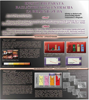
Dr Savic-Stankovic defended her MSc thesis in a viva but also presented the highlights of her research at the School of Dentistry Anniversary in June 2009. Click on the image to enlarge the poster. Since the poster is in Serbian, the abstract in English is given below. Copyright belongs to Dr Savic-Stankovic. For further contact, please use the email address savic.t@nadlanu.com


The effect of different concentrations of bleaching agents on enamel properties in vitro and clinical efficiency of the „walking bleach“ technique

Dr Tatjana Savic-Stankovic BDS, MSc

Introduction. Bleaching results in slow transformation of organic substances into chemical intermediary products which are lighter than the original. Bleaching efficiency is related to the cause of tooth discoloration.

Aims. To evaluate changes in enamel microhardness and morphological aspects of enamel after treatment with different concentrations of bleaching agents. The aim of the clinical study was to evaluate the bleaching efficiency with regard to the cause of discoloration: trauma, necrosis, endo-sealer or unknown.

Materials and Methods. Twenty freshly extracted sound human molars were treated with 10%, 20% and 35% carbamide peroxide and 38% hydrogen peroxide (Opalescence gel, Ultradent. Pro.) in clinically recommended intervals. Knoop microhardness measurements were performed at baseline and 8 hours, 21 days and 3 weeks post-treatment. SEM was used to evaluate morphological changes in enamel post-treatment.

Forty non-vital central incisors were bleached using sodium perborate and hydrogen peroxide mixture which was changed weekly. Based on patients' dental history and clinical examination, discolored teeth were allocated to four groups depending on the cause of discoloration: trauma, necrosis, endo-sealer and unknown. Aesthetic results of the applied “walking bleach” technique were evaluated before and after treatment using the standardized colour-coded key for colour comparison.

Results. No statistically significant differences and no changes in enamel surface morphology were observed between 10% and 20 % carbamide peroxide groups. A significant decrease in enamel microhardness was found in 35% carbamide peroxide and 38% hydrogen peroxide groups. Mild and moderate changes in enamel surface morphology were observed in the group treated with 35% carbamide peroxide whilst substantial changes were found in the group treated with 38% hydrogen peroxide.

The shortest treatment (15.7 days) and greatest number of shades before and after bleaching were found in patients whose aetiological factor for discoloration was „trauma”. Bleaching efficiency decreased with age. No correlation was found between bleaching efficiency and the initial shade.

Conclusions. Increasing concentrations of tooth-bleaching agents resulted in lower enamel microhardness values and more profound changes in enamel surface morphology. The cause of tooth discoloration had a significant effect on the clinical efficiency of the "walking bleach" technique.

Keywords: dental materials, tooth bleaching, tooth whitening, carbamide peroxide, hydrogen peroxide, enamel, clinical trial, walking bleach

Monday 7 September 2009

IADR-CED with NOF & ID Conference, Munich, Sept 9-12, 2009


The 44th Conference of the Central European, Scandinavian and Israeli Divisions of IADR


The scientific programme includes several oral sessions, poster presentations and workshops. There will be several dental materials symposia:
  • Zirconiumdioxide restorations and reasons for failures
  • New composite formulations
  • Self adhesive cements
More than 400 studies will be presented at the Conference. Complete programme and author index can be found HERE.

Two of my students, Ana Ergic and Dejan Nedeljkovic, will present our study entitled "Surface characterisation of resin-based composite materials using atomic force microscopy". The poster will be uploaded to this blog after the conference.

Sunday 6 September 2009

Journal of Dentistry: Top 10 Hottest articles


The list of Top 10 Hottest articles, most frequently downloaded from Journal of Dentistry is regularly published on this journal's website. The latest list can be found HERE.

Eight out of ten most downloaded articles are related to dental materials. The single most popular article is the review of the effect of polyphenols on oral health and disease. The most popular field represented by the greatest number of articles (4) in the Top 10 seems to be the bleaching of teeth. The most popular research article is a study on the effect of different adhesive systems and laser treatment on the shear bond strength of bleached enamel.

Journal of Dentistry is a SCI journal, published monthly, with the current impact factor of 2.033.


Thursday 3 September 2009

Latest jobs in dental materials science

PhD Studentship

The Impact Of Titanium Accumulation On Peri-Prosthetic Soft Tissue Outcomes
School of Dentistry , College of Medical and Dental Sciences — University of Birmingham

Application deadline: 07 Sep 2009

************************************************************************************
I'd like to add a personal remark that this post offers an opportunity to work with one of the most competent dental material scientists, Dr Owen Addison, in an intellectually stimulating scientific environment at the University of Birmingham School of Dentistry.

************************************************************************************
NB. I'm proud to announce that more jobs related to dental materials will be posted on this blog with permission from jobs.ac.uk. For information on other jobs, visit www.jobs.ac.uk and check out blogs by their resident bloggers who share experiences of jobseeking, academic life and more.

BSDR Conference in Glasgow

As previously announced, the BSDR Conference is taking place in Glasgow. The Santini Miletic Research Group presented a study entitled "The ratio of carbon-carbon double bonds in different BisGMA/HEMA mixtures". Click on the image.
Prior to the poster session, I attended the Ceramics session chaired by professor Richard Van Noort. Several very interesting studies were presented regarding CAD-CAM ceramic strength, ceramic reinforcements, fluoride-containing bioactive glasses, leucite glass-ceramic crystallisation, coating materials for zirconia ceramics and wear quantification using profilometry. It was quite impressive to see the very high standard of research carried out at various universities in the UK.

Sponsored by 3M ESPE, the Dental Materials Group Symposium was held during the afternoon session. After the opening remarks by Dr Garry Fleming and Professor R. Van Noort, lectures were given by Dr Rainer Guggenberger (The chemistry of new resin systems), Professor David Watts (The measurement of shrinkage and contraction stress), Professor Tim Watson (The quality of adhesion) and Professor Trevor Burke (Early thoughts of clinical experience using the novel silorane-based composite material). The lectures increased our knowledge on various aspects of resin-based composites and the silorane-based material in particular. The 'non-shrink' resin composites are still not a reality but dental technology has made substantial improvements towards this goal.

Sunday 30 August 2009

Publishing papers: initial observations as an author and a referee

Three basic rules for referees

I've published 9 papers in peer-reviewed dental journals in the last 2 years and have refereed half a dozen papers regarding dental materials, adhesives, resin-based composites, adhesive cements, adhesion, marginal adaptation... Since there's no 'school' or 'course' for referees that I'm aware of, I've started developing my own style. Judging from other people's reviews of my papers, this seems to be the case with most referees out there. I know that the following are very basic rules, but sometimes, even such basic rules are not followed.

Though editors put a lot of effort in finding the right referees for a particular paper, it's possible that a paper ends up in wrong hands, with someone whose field of expertise has almost nothing to do with the subject of the paper. I've noticed very 'interesting' remarks regarding one of my papers indicating that the referee wasn't an expert in the field he/she was referring to. Obviuosly, nowadays a lot of studies are multidisciplinary and this makes it even more difficult for editors to find competent referees. A potential referee may be an expert regarding only a part of a particular study. It's only fair to comment on that part and indicate this to the editor so that a competent person is invited to comment on the other part(s).

So, rule No.1: accept an invitation to review a paper only if you're an expert in the field or at least a part of it and indicate this to the editor. Decline review if the subject of the paper is not your area of expertise.

Another thing I've noticed with people refereeing my papers is that this process sometimes takes ages. Though some journals have taken steps to ensure speedy review within 3-6 weeks, with some journals this may take even more than 6 months. This is simply unacceptable. Being in a referee's position myself, I've realised that even 3 weeks is more than enough to complete a review. Of course, I'm well aware of the fact that referees are very busy and have many, many other things to do. But so are the authors and referees should respect that. It's unfair to keep someone's paper 'under review' for more than a month.

So, rule No.2: accept an invitation to review a paper only if you can complete the review in less than 3 weeks.

Also, I've noticed that there are people who just don't care about the paper they're reviewing. They tend to write and overall impression of the paper without going into specific details. This is not helpful even if the paper is good and especially if it needs to be improved. It's almost like they just skimmed through the paper. On the other hand, there are people who write extensive reviews and I find these extremely helpful.

So, rule No.3: be fully committed to reviewing a paper. Go into minute details when writing a review.

Keywords: dental materials, research, publishing papers, review process.

Tuesday 25 August 2009

Santini Miletic Research Group: new website


The new website of the Santini Miletic Research Group is up and running. Please visit us at http://www.santinimiletic.com/


In addition to the short CVs of group members, there is a list of selected papers published recently in international peer-reviewed journals. There is also information on research facilities available to the Group and various methods of knowledge transfer.

Friday 21 August 2009

Up-coming event

British Society for Dental Research Conference, Glasgow, Sept 1-4, 2009.

The keynote speakers are Professor Angus Walls, Professor of Restorative Dentistry, School of Dental Sciences, Newcastle University and Professor Iain McInnes, Professor of Experimental Medicine, Division of Immunology, Infection and Inflammation, Faculty of Medicine, University of Glasgow.

Professor Angus Walls' lecture - "The ageing population, opportunity or threat?"
Professor McInnes' lecture - "Immune complexity to novel therapy - promises for a new decade?"

The following symposia are scheduled:

- Non-shrink resin composites: Dream or reality?
Aim: This symposium examine current developments of resin-based restoratives, indicating the advantages and disadvantages of resin types routinely and review the most recent advancements in resin technology.
Speakers:
Professor Ric van Noort, University of Sheffield: Opening remarks
Dr Rainer Guggenberger, 3M/ESPE: The chemistry of new resin systems
Dr N Silikas University of Manchester: Measurement of shrinkage and contraction stress
Professor Tim Watson Kings College London: Quality of adhesion
Professor Trevor Burke University of Birmingham: Early thoughts of clinical experience

- Novel biomaterials - can the microenvironment be improved ?

- Childsmile a Scottish oral health programme for Scottish children: collaborations and innovative evaluations

- The oral microbiota and the link with systemic disease

A total of 228 studies will be presented at the conference and the complete programme can be downloaded from HERE.

The Santini Miletic Research Group will present a study entitled "The ratio of carbon-carbon double bonds in different BisGMA/HEMA mixtures". The poster will be uploaded to this blog after the conference.

Thursday 20 August 2009

IADR introduces official social networking platforms

Official IADR communities have been created on Facebook, LinkedIn and Flickr to enhance communication between IADR members. Anyone can join. It's free. Click on each logo below for more.


Wednesday 19 August 2009

One-step self-etch adhesive, Adhese One F

A new one-step self-etch adhesive, Adhese One F, manufactured by Ivoclar Vivadent has been sent to the Santini Miletic Research Group for scientific evaluation. The adhesive is based on previously developed Adhese One with the inclusion of potassium fluoride which is reported to act as a fluoride releasing agent. The manufacturer's internal data state that there is a cumulative fluoride release over a 6 day period.

Micro-Raman spectroscopic studies will be conducted to evaluate the ratio of carbon-carbon double bonds (RDB) of Adhese One F under different curing conditions. Furthermore, the adhesive-dentine interface will be characterised in terms of dentine demineralisation and adhesive penetration and the RDB across this interface.

A previous study has shown significantly lower RDB values for Adhese One in both the adhesive and the hybrid layer compared to Excite (etch-and-rinse) and Adhese (2-step self-etch). In another study, Adhese One produced a thinner hybrid layer compared to G Bond (1-step self-etch), Filtek Silorane adhesive system (2-step self-etch) and Excite

Santini A, Miletic V. Quantitative micro-Raman assessment of dentine demineralization, adhesive penetration, and degree of conversion of three dentine bonding systems. Eur J Oral Sci 2008;116(2):177-83. Abstract Full text available upon request.

Santini A, Miletic V. Comparison of the hybrid layer formed by Silorane adhesive, one-step self-etch and etch and rinse systems using confocal micro-Raman spectroscopy and SEM. J Dent 2008;36(9):683-91. Abstract Full text available upon request.