Social Icons

Pages

Thursday, 28 July 2011

Clinical reproducibility of three electronic apex locators

Though it is not quite a dental materials subject, I have been involved in clinical testing of electronic apex locators with my colleagues at the University of Belgrade School of Dentistry. This paper has been published in the August issue of the International Endodontic Journal and can be found here. If you have trouble getting access, please email me.
**********************************************************************************
Miletic V, Beljic-Ivanovic K, Ivanovic V. Clinical reproducibility of three electronic apex locators. International Endodontic Journal, 44, 769–776, 2011.

Abstract

Aim  To compare the reproducibility of three electronic apex locators (EALs), Dentaport ZX, RomiApex A-15 and Raypex 5, under clinical conditions.
Methodology  Forty-eight root canals of incisors, canines and premolars with or without radiographically confirmed periapical lesions required root canal treatment in 42 patients. In each root canal, all three EALs were used to determine the working length (WL) that was defined as the zero reading and indicated by ‘Apex’, ‘0.0’ or ‘red square’ markings on the EAL display. A new K-file of the same size was used for each measurement. The file length was fixed with a rubber stop and measured to an accuracy of 0.01 mm. Measurements were undertaken by two calibrated operators. Differences in zero readings between the three EALs in the same root canal were statistically analysed using paired t-tests with the Bonferroni correction, Bland–Altman plot and Linn’s concordance correlation coefficients at α = 0.05.
Results  Mean and standard deviation values measured by the three EALs showed no statistically significant differences. Identical readings by all three EALs were found in 10.4% of root canals. Forty-three per cent of readings differed by less than ±0.5 mm and 31.3% exceeded a difference of ±1 mm.
Conclusions  The clinical reproducibility of Dentaport ZX, RomiApex A-15 and Raypex 5 was confirmed with the majority of readings within the ±1.0 mm range. However, the small number of identical zero readings suggests that EALs are not reliable as the sole means of WL determination under clinical conditions.

Saturday, 18 June 2011

Upcoming event: CED IADR 2011

The 45th Meeting of the Continental European Division (CED) of IADR, organized together with the Scandinavian Division (NOF) will be held in Budapest, Hungary, from August 31 till September 3, 2011. Basic sciences and clinical topics, such as dental materials, cariology, implantology, periodonontology, oral medicine, tissue engineering, craniofacial biology, salivary research etc. will be covered by plenary lectures, symposia, oral and poster presentation sessions etc.

Scientific symposia on dental materials will cover the following subjects:
  1. Adhesive technology
  2. Needs and requirements of dental materials in combating caries 
  3. Biomaterials in periodontal and implant surgery
  4. Aesthetic and mechanical aspects of composite materials
  5. Implant restoration
  6. Nano biomaterials in regenerative dentistry
  7. Novel tri-calcium silicate-based dentine substitute
I will present a digital image correlation study on composite shrinkage using a two-camera system which allows 3D measurement of strains and displacements. This study is a result of strengthening research ties between my team from the School of Dentistry and colleagues from the Faculty of Mechanical Engineering, University of Belgrade, Serbia. The abstract of this study is here. Also, other studies that will be presented during the same session on Composites, Shrinkage, Curing, and Fracture Toughness may be found here.

For more information on the scientific programme and registration for the 45th IADR CED&NOF conference please visit the official CED-IADR2011 website.

    Tuesday, 31 May 2011

    Free e-learning resources on glass ionomers

    Vivalearning.com is a popular e-learning resource featuring lots of webinars on various dental topics. All webinars are free and CE credited! All webinars premier as live presentations and then are accessible on demand.

    The Wonderful World of Glass Ionomer in Clinical Dentistry
    Presentedby Dr Jeff Brucia

    LIVE ON (click here to register and reserve seat)
    Thursday June 6, 2011 8:00 PM ET / 5:00 PM PT
    Friday June 7, 2011 0:00 AM UTC (former GMT)
    CE Credits: 1

    From Vivalearning.com: "A comprehensive discussion of the ever-changing world of Glass Ionomers will address the following questions. Do they still cause sensitivity? Are they any more aesthetic? Are they strong enough? How and where should they be used? Do they adhere to tooth structure? This presentation is a must for any practitioner that is considering an operative procedure in a less than ideal clinical situation."


    Modern Glass Ionomers Used as Liners in the Composite Resin Sandwich Technique
    Presented by Greg Gillespie DDS, Lou Graham DDS and Mark A. Latta DMD, MS
    CE Credits: 1
    On Demand (click here to register and access class)

    This presentation was released on January 7, 2011. It is now in the form of a downloadable PDF file. The authors give a short history of glass ionomers, explain the concept of the "sandwich" technique and provide several cases to illustrate this clinical procedure.

    Sunday, 22 May 2011

    How to measure the depth of cure of composites according to ISO 4049?

    The ISO4049 standard explains in detail how the depth of cure is measured and what is minimum depth that composites must have in order to comply with this standard. This simple procedure does not require sophisticated equipment and may be done in every dental office. It allows testing and comparison of materials and light curing units. Even if there is a radiometer to check the light intensity, it is recommended to measure the actual thickness of the composite cured by a a particular light curing unit.

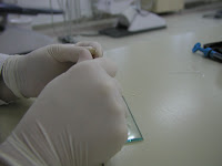
    Here is what we need:
    1. composite
    2. light curing unit
    3. cylindrical moulds (6 mm thick and 4-5 mm in diameter), originally it should be stainless steel, but plastic straws cut into moulds of this size may be used as well
    4. glass slab
    5. Mylar strips
    6. plastic filling instrument
    7. spatula or scalpel
    And here is the step-by-step procedure:

    1. Place the mould on the glass slab and fill it with composite.


    2.  Place the Mylar strip on top of the composite.


    3. Light-cure the composite according the manufacturer's instructions (i.e. 40 s using a conventional or 20 s using a high-power halogen or LED light).


    4. Discard the Mylar strip and remove the cured material from the mould.


    5. Peel off the uncured material from the bottom side of the sample using the spatula or scalpel.


    6. Measure the remaining thickness of the sample and divide this number by two. The ISO 4049 standard requires that the result should be at least 1.5 mm for non-opaque shades and 0.5 mm for opaque shades.

    Tuesday, 26 April 2011

    Free webinar on posterior composites

    Posterior Composites: Improving Esthetics and Increasing Simplicity 
    Presenter: Dr. Greg Gillespie

    (CE credits 1)

    Date Wednesday 27 April 2001
    Time 7:00 pm ET / 6:00 pm CT / 4:00 pm PT / 11 pm UTC (former GMT)


    This webinar is sponsored by GC America.
    From the official website: "In this webinar, Dr. Gillespie will review adhesive protocols that will help eliminate post-operative sensitivity and increase bond strengths. Dr. Gillespie will also highlight the latest advancements in composite resins regarding shrinkage and esthetics with one shade placement."

    It is necessary to register and reserve a seat. If you miss the live webinar, it will soon be among On demand webinars. They are all free and may be accessed at any time.

    Friday, 8 April 2011

    Glass Ionomer - Composite "sandwich" technique: when is the time to etch?

    Glass-ionomer cements (GIs) are still the only true self-adhesive materials forming the chemical bond with tooth tissues. Despite the traditional classification to types of GIs, current scientific literature is dominated by a simpler and yet more informative classification to conventional and resin-modified GIs. This indicates information about materials' chemical composition, curing mode and clinical application steps.

    The difference between conventional and resin-modified GIs is in the organic resin monomers added to the latter formula which enables prompt light-curing of the material using halogen or LED units. Light curing of resin creates favourable micro-environment for the conventional acid-base reaction between polyacrilic acid and glass particles. Improvements in material composition have led to improved mechanical properties although GIs are still inferior compared to resin-based composites. On the other hand, sensitivity to water imbalance, characteristic for early GIs, has been largely overcome in modern GIs both conventional and resin-modified. More information about GIs, their composition, properties and indications may be found in an excellent review article by Hewlett and Mount, published in 2003. [Full text]

    One of the indications for GIs is the so-called "sandwich" technique with composite materials for large restorations on both vital and endodontically treated teeth. According to manufacturers' instructions both conventional and resin-modified GIs may be used for this purpose. Though it is widely known that early GIs were sensitive to water imbalance during setting, there is a certain controversy regarding this issue with current GIs. Due to this controversy, a clinical dilemma exists among dental practitioners when using GIs in combination with total-etch adhesives prior to composite placement. This dilemma is not about the acid but rather water rinsing afterwards. Simply, some practitioners are not convinced that GIs should be exposed to water so early after the setting (e.g. 3 minutes for FUJI IX GP Fast) or immediately after light curing of resin-modified GIs.

    The manufacturer recommends the following protocol (Figure 1):
    Figure 1. GC Europe recommends enamel etching after the placement of GI intermediary layer. (1) Old amalgam restoration; (2) Cavity preparation; (3) Conditioning; (4) Application of GI ; (5) GI layer ready; (6) Enamel etching; (7) Application of adhesive; (8) Light curing and (9)-(12) Placement of composite.
    An alternative protocol suggested by some dental practitioners (Figure 2):

    Figure 2. Enamel etching prior to the placement of GI. From left to right upper row: Cavity preparation; Conditioning; Enamel etching. From left to right bottom row: Application of GI; Application of adhesive; Final composite restoration.


    The second approach does eliminate the possible adverse effect of water during acid rinsing. However, acid etching and rinsing prior to GI placement bears an inherent weakness - this approach requires impeccable precision. Dentin should not be etched if GI is to be placed since the mineral component required for chemical bonding will be lost. On the other hand, if adhesive is to be placed on dentin as well as on enamel, dentin should also be etched for proper micro-mechanical bonding of adhesive resin.

    If one does not want to follow manufacturer's instructions and acid etch after the placement of the GI layer, then they should consider the use of self-etch adhesives instead of total-etch systems. One-step self-etch adhesives have shown inferior results regarding bond strength to dentin and enamel, degree of conversion, thickness of the hybrid layer, the quality of resin tags etc. On the other hand, current 2-step self-etch adhesives have shown satisfactory clinical and laboratory properties in a number of studies and are recommended as an alternative to total-etch adhesive systems.

    In my practice, I always follow manufacturer's recommendations. In this case, I use resin-modified GIs for the "sandwich" technique and acid etch enamel after light curing of the GI intermediary layer.

    Friday, 25 March 2011

    IADR Toshio Nakao Fellowship

    The 89th General Session of the International Association for Dental Research was held last week in San Diego, CA, USA. I was officially awarded the IADR Toshio Nakao Fellowship during the opening ceremony. This Fellowship is generously supported by GC Corporation with the intention to "allow a young investigator to obtain training and experience in dental materials science at a center of excellence". In my case, this means six months post-doctoral research at the University of Edinburgh, Great Britain, commencing in June 2011. It was truly an honour to be among the distinguished scientists whose work has been recognized by the IADR, the leading international organisation in dental research. I am grateful to the IADR and GC Corporation for support. This Fellowship will help me continue research on dental adhesives, particularly monomer to polymer conversion and hybridisation of dentine.

    Tuesday, 8 March 2011

    A review of SEM and TEM studies on the hybridisation of dentine

    Professors Ario Santini and Egle Milia and Dr Vesna Miletic, members of the Santini Miletic Research Group, published a chapter on dentine hybridisation in the international peer-reviewed book Microscopy: Science, Technology, Applications and Education, edited by A. Mendez-Vilas and J. Diaz and published by Formatex (www.formatex.org). This is the 4th book in the Microscopy series. The chapter is a review of SEM and TEM studies on the hybridisation of dentine with additional findings on the subject using micro-Raman spectroscopy. The chapter can be downloaded free of charge from the publisher's website
    http://www.formatex.org/microscopy4/chapters1.html (simply scroll down to Santini, Milia and Miletic).

    Santini A, Egle M and Miletic V. A review of SEM and TEM studies on the hybridisation of dentine.
    In: Microscopy: Science, Technology, Applications and Education.
    Editors: Mendez-Vilas A, Diaz J. Microscopy series No. 4, Volume 1, pages 256-268.       
    Publisher: FORMATEX, Badajoz, Spain, 2011.
    ISBN-13: 978-84-614-6189-9

    Abstract

    Current opinion is that the hybridisation of dentine is the principal, though not the exclusive, mode of adhesion of restorations to tooth tissue. Hybrid layer formation is achieved by resin infiltration of acid-etched dentine. This layer provides micromechanical retention for resin composite restorations.

    The vast literature on the development of bonding systems is summarised and the differences in the hybrid layer formation are reviewed with specific attention to SEM and TEM studies.

    It is concluded that more recently marketed adhesives with simplified application procedures are less successful compared to conventional total-etch adhesives.

    **********************************************************************************

    Visit santinimiletic.com for more references of our research group.

    Tuesday, 15 February 2011

    Water sorption and solubility of resin-based composites

    Interaction of resin-based composites with water is a continuous process from the early stages of composite placement. Water plays an important role in the long-term stability of composite fillings and may induce hygroscopic expansion of the material, hydrolytic degradation of intra- and intermolecular bonds within the resin matrix and at the resin-filler interface, plasticization of polymer chains, elution of leachable substances and reduction in mechanical properties.

    The importance of composite-water interaction has been acknowledged in the ISO standard 4049 which states the maximum values for water sorption and concurrent solubility for resin-based materials (composites and cements). In order to comply with this ISO standard, resin-based materials must have  water sorption and solubility values equal or lower than 40 micrograms per cubic milimetre (sorption) and 7.5 micrograms per cubic milimetre (solubility) for specimens 15 mm in diameter and 2 mm thick.

    Water sorption and solubility values are based on mass changes of the samples before (m1) and after immersion (m2) in water and after dessication (m3) until constant mass is achieved.  Mass changes m2-m3 are divided by sample volume to calculate water sorption and m1-m3 divided by sample volume give solubility values. 

    Recent papers* published in Dental Materials investigated water sorption and solubility and hygroscopic dimensional changes of several resin-based composites:

    low-shrinkage Filtek Silorane
    universal Gradia Kalore
    micro-hybrid Gradia Direct Anterior and Posterior and
    self-adhering flowable Vertise Flow.

    After 150 days of storage in de-ionized water, the lowest sorption of about 13 μg/mm³ was found for Filtek Silorane and the greatest of about 72 μg/mm³ was found for Vertise Flow. Vertise Flow also showed the greatest solubility of about 16 μg/mm³ whereas other materials showed either negative values (Filtek Silorane and Gradia Kalore) or values below 4 μg/mm³. The authors suggested that the negative solubility values for Filtek Silorane and Gradia Kalore meant that the dessication was not sufficient or that some water was irreversibly bound to the resin matrix.

    Hygroscopic dimensional expansion as the result of water sorption over the 150-day period was lowest for Filtek Silorane (about 0.7%) and highest for Vertise Flow (about 4.8%) whereas the values for Gradia composites were between 1.5 and 2%. Hygroscopic expansion may compensate to a certain extent polymerization shrinkage which was found to be 0.99% for  Filtek Silorane, 1.7-2.4% for Gradia composites and  4.4% for Vertise Flow. However, this expansion occurs over a much slower time scale than shrinkage and its effect on the clinical performance of resin-based composite is yet to be determined.

    The greatest stability in the aqueous environment found for Filtek Silorane may be explained by the hydrophobic siloxane and low-shrinkage ring-opening oxirane units of the silorane monomer. Furthermore, cationic polymerization is relatively oxigen-insensitive with the potential of reaching higher degree of conversion than methacrylate-based composites.

    On the other hand, aqueous instability of Vertise Flow was attributed to the hydrophilic monomer, GPDM, which is responsible for the self-adhesive property of Vertise Flow but also seems to attract more water uptake by the resin matrix compared to other resin-based composites.


    * Wei YJ, Silikas N, Zhang ZT, Watts DC. Hygroscopic dimensional changes of self-adhering and new resin-matrix composites during water sorption/desorption cycles. Dent Mater. 2011 Mar;27(3):259-66.


    [Reprints of the cited papers may be obtained from the corresponding authors]

    Click here for more on Vertise Flow.
     

    Wednesday, 2 February 2011

    Monomer elution from nanohybrid and ormocer-based composites

    Recently, a paper on monomer elution from resin-based composites by my research team has been accepted for publication in the leading peer-reviewed journal on dental materials. The paper is available online under "Articles in press" and awaits publication in the paper format of Dental Materials. For reprints, you may contact me at vesna.miletic@gmail.com 


    Abstract

    Dent Mater. 2010 Dec 17. [Epub ahead of print]

    Monomer elution from nanohybrid and ormocer-based composites cured with different light sources

    Manojlovic D, Radisic M, Vasiljevic T, Zivkovic S, Lausevic M, Miletic V.

    University of Belgrade, School of Dentistry, Department of Restorative Dentistry
    and Endodontics, Rankeova 4, 11000 Belgrade, Serbia.


    OBJECTIVES: To study monomer elution from four resin-based composites (RBCs)cured with different light sources.  
    METHODS: Twenty-eight premolars were randomlyallocated to four groups. Standardized cavities were prepared and restored with ananohybrid (Filtek Supreme XT or Tetric EvoCeram), an ormocer (Admira) or a microhybrid RBC (Filtek Z250) which served as control. Buccal restorations were cured with a halogen and oral restorations with an LED light-curing unit. Elution of diurethane dimethacrylate (UDMA), Bisphenol A diglycidylether methacrylate (BisGMA), triethylene glycol dimethacrylate (TEGDMA) and 2-hydroxyethyl methacrylate (HEMA) was analyzed using high-performance liquid chromatography (HPLC) 1h to 28 days post-immersion in 75% ethanol. Data were analyzed using multivariate and repeated measures analysis of variance (α=0.05).  
    RESULTS: The greatest elution of UDMA and BisGMA occurred from Tetric EvoCeram and the least from Filtek Z250 (p<0.05). LED and halogen light-curing units gave similar results for all RBCs (p>0.05) except Tetric EvoCeram which showed greater elution for the LED unit (p<0.05). TEGDMA was below the limit of quantification. HEMA eluted in similar concentrations from Filtek Supreme and Tetric EvoCeram (p>0.05).  
    SIGNIFICANCE: The two nanohybrid RBCs eluted more cross-linking monomers than the ormocer and the control microhybrid RBC. Continuous elution over 28 days indicates that RBCs act as a chronic source of monomers in clinical  conditions. Light source may affect monomer elution since differences were found  for one out of four RBCs. Mathematical models for elution kinetics of UDMA and BisGMA indicated two elution mechanisms.

    Monday, 24 January 2011

    News from jobs.ac.uk

    PhD Studentship (full time, 3 years)

    Toxicological Studies of Endocrine Disrupting Chemicals in Dental Patients
    Peninsula College Of Medicine And Dentistry Graduate School


    This studentship aims to investigate the baseline and post-treatment urine and saliva concentrations of Bisphenol A and phthalate plasticisers in paediatric and adult patient populations entering Peninsula Dental School (PDS) Clinics.

    The post will involve chromatographic (LC-MS/MS) analysis of biological samples and potentially, advanced mathematical modelling of data obtained. We are therefore looking for someone with a science background (BDS, BMBS, BSc Biomedical Sciences/Biology/Pharmacology/Chemistry or similar) with an aptitude for mathematics.

    Stipend:  £13,590 
    UK/EU candidates only
    Closing date: 21 February 2011

    For more information, click here

    Saturday, 15 January 2011

    Dental Materials Conferences 2011

    89th General Session & Exhibition of the IADR
    40th Annual Meeting of the AADR
    35th Annual Meeting of the CADR
    March 16-19, 2011
    San Diego, California, USA
    More information on the IADR website. Click here for the online scientific programme. More than 4000 abstracts are expected to be featured at the conference. Traditionally, the greatest number of sessions will cover many aspects of dental materials, from chemistry and properties to clinical performance.

    45th Meeting of the Continental European Division (CED) and Scandinavian Division (NOF) of the IADR
    August 31-September 3, 2011
    Budapest, Hungary
    More information on the IADR CED website. Deadline for abstract submission is March 25, 2011. Click here to submit your abstract. The CED-IADR will provide considerable support to young researchers through as many travel stipends as possible.

    21st European Dental Materials Conference
    August 24-26, 2011
    Turku, Finland
    “Current trends in moving toward non-metallic materials”
    More information on the conference website. Click here for the tentative scientific programme. Dental Materials Summer School will be organized prior to the conference.

    2011 Conference of the Academy of Dental Materials
    October 13-15, 2011.
    Bahia, Brazil
    For more information, click here. Topics at this conference: What is the future? When to market? Where will the material be used?

    International Dental Materials Congress
    May 27-29, 2011
    Seoul, Korea
    "Moving Dental Materials from the Laboratory to the Clinic"
    Visit the Congress website for more information. Click here for the scientific programme.