Social Icons

Pages

Showing posts with label Clinical practice. Show all posts

Sunday 22 May 2011

How to measure the depth of cure of composites according to ISO 4049?

The ISO4049 standard explains in detail how the depth of cure is measured and what is minimum depth that composites must have in order to comply with this standard. This simple procedure does not require sophisticated equipment and may be done in every dental office. It allows testing and comparison of materials and light curing units. Even if there is a radiometer to check the light intensity, it is recommended to measure the actual thickness of the composite cured by a a particular light curing unit.

Here is what we need:
  1. composite
  2. light curing unit
  3. cylindrical moulds (6 mm thick and 4-5 mm in diameter), originally it should be stainless steel, but plastic straws cut into moulds of this size may be used as well
  4. glass slab
  5. Mylar strips
  6. plastic filling instrument
  7. spatula or scalpel
And here is the step-by-step procedure:

1. Place the mould on the glass slab and fill it with composite.


2.  Place the Mylar strip on top of the composite.


3. Light-cure the composite according the manufacturer's instructions (i.e. 40 s using a conventional or 20 s using a high-power halogen or LED light).


4. Discard the Mylar strip and remove the cured material from the mould.


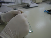
5. Peel off the uncured material from the bottom side of the sample using the spatula or scalpel.


6. Measure the remaining thickness of the sample and divide this number by two. The ISO 4049 standard requires that the result should be at least 1.5 mm for non-opaque shades and 0.5 mm for opaque shades.

Friday 8 April 2011

Glass Ionomer - Composite "sandwich" technique: when is the time to etch?

Glass-ionomer cements (GIs) are still the only true self-adhesive materials forming the chemical bond with tooth tissues. Despite the traditional classification to types of GIs, current scientific literature is dominated by a simpler and yet more informative classification to conventional and resin-modified GIs. This indicates information about materials' chemical composition, curing mode and clinical application steps.

The difference between conventional and resin-modified GIs is in the organic resin monomers added to the latter formula which enables prompt light-curing of the material using halogen or LED units. Light curing of resin creates favourable micro-environment for the conventional acid-base reaction between polyacrilic acid and glass particles. Improvements in material composition have led to improved mechanical properties although GIs are still inferior compared to resin-based composites. On the other hand, sensitivity to water imbalance, characteristic for early GIs, has been largely overcome in modern GIs both conventional and resin-modified. More information about GIs, their composition, properties and indications may be found in an excellent review article by Hewlett and Mount, published in 2003. [Full text]

One of the indications for GIs is the so-called "sandwich" technique with composite materials for large restorations on both vital and endodontically treated teeth. According to manufacturers' instructions both conventional and resin-modified GIs may be used for this purpose. Though it is widely known that early GIs were sensitive to water imbalance during setting, there is a certain controversy regarding this issue with current GIs. Due to this controversy, a clinical dilemma exists among dental practitioners when using GIs in combination with total-etch adhesives prior to composite placement. This dilemma is not about the acid but rather water rinsing afterwards. Simply, some practitioners are not convinced that GIs should be exposed to water so early after the setting (e.g. 3 minutes for FUJI IX GP Fast) or immediately after light curing of resin-modified GIs.

The manufacturer recommends the following protocol (Figure 1):
Figure 1. GC Europe recommends enamel etching after the placement of GI intermediary layer. (1) Old amalgam restoration; (2) Cavity preparation; (3) Conditioning; (4) Application of GI ; (5) GI layer ready; (6) Enamel etching; (7) Application of adhesive; (8) Light curing and (9)-(12) Placement of composite.
An alternative protocol suggested by some dental practitioners (Figure 2):

Figure 2. Enamel etching prior to the placement of GI. From left to right upper row: Cavity preparation; Conditioning; Enamel etching. From left to right bottom row: Application of GI; Application of adhesive; Final composite restoration.


The second approach does eliminate the possible adverse effect of water during acid rinsing. However, acid etching and rinsing prior to GI placement bears an inherent weakness - this approach requires impeccable precision. Dentin should not be etched if GI is to be placed since the mineral component required for chemical bonding will be lost. On the other hand, if adhesive is to be placed on dentin as well as on enamel, dentin should also be etched for proper micro-mechanical bonding of adhesive resin.

If one does not want to follow manufacturer's instructions and acid etch after the placement of the GI layer, then they should consider the use of self-etch adhesives instead of total-etch systems. One-step self-etch adhesives have shown inferior results regarding bond strength to dentin and enamel, degree of conversion, thickness of the hybrid layer, the quality of resin tags etc. On the other hand, current 2-step self-etch adhesives have shown satisfactory clinical and laboratory properties in a number of studies and are recommended as an alternative to total-etch adhesive systems.

In my practice, I always follow manufacturer's recommendations. In this case, I use resin-modified GIs for the "sandwich" technique and acid etch enamel after light curing of the GI intermediary layer.

Sunday 26 December 2010

Clinical evaluation of dental restorative materials - Part III: FDI criteria

In 2007, recommendations for conducting clinical trials approved by the FDI were published in several peer-reviewed journals. These recommendations not only addressed designing protocols for clinical trials but also challenged the Ryge criteria with an in-depth discussion of  clinical evaluation criteria and suggested a new approach in clinical evaluation of dental restorative materials and operative techniques.

In August 2010, an update of the "FDI clinical criteria for the evaluation of direct and indirect restorations" was published  by Hickel et al. in the Journal of Adhesive Dentistry and Clinical Oral Investigations. A lot of clinical examples were presented in the paper to illustrate various ratings.

The new FDI criteria set a different background for the evaluation of dental restorations by introducing 3 groups of criteria: esthetic, functional and biological. Each of these groups has subgroups with 16 evaluation criteria in total. These are:

Esthetic criteria
  1. Surface luster
  2. Staining: (a) surface and (b) margin
  3. Color match and translucency
  4. Esthetic anatomical form
Functional criteria
  1. Fracture of material and retention
  2. Marginal adaptation
  3. Occlusal contour and wear 
  4. Approximal anatomical form: (a) contact point and (b) contour
  5. Radiographic examination, where applicable
  6. Patient's view
Biological criteria
  1. Postoperative sensitivity and tooth vitality
  2. Recurrence of caries, erosion, abfraction
  3. Tooth integrity
  4. Periodontal response
  5. Adjacent mucosa
  6. Oral and general health
For all three groups, the following gradings are used for evaluation:
  1. Clinically excellent/very good
  2. Clinically good
  3. Clinically sufficient/satisfactory
  4. Clinically unsatisfactory
  5. Clinically poor
When judging a dental restoration using the FDI criteria, the score for each group is dictated by the most severe grading among the criteria for that particular group. Similarly, the overall score is determined by the worst grading among the groups. For example, if the functional criteria are unacceptable, the overall score is unacceptable. Detailed description of each grading is given in the previously mentioned paper: "FDI World Dental Federation - Clinical Criteria for the evaluation of direct and indirect restorations".

It is not mandatory to apply all of the FDI criteria in each study. In each particular study, the examiners should determine which criteria match their intended purposes best.

Gradings for the FDI criteria are substantially more detailed and sensitive than the Cvar and Ryge criteria and their modifications suggested by other authors. These detailed gradings challenge the training and calibration procedure of the examiners. To allow an easier and more efficient training, reduced variability in judgment and greater coherence in multi-centric studies, an online calibration system was established at www.e-calib.info. It is emphasized by the authors of the FDI criteria that the e-calibration system does not replace the clinical setting but shortens clinical training significantly.

Beside their use in clinical trials, the FDI criteria are recommended for quality assessment of restorations by general dental practitioners in their everyday practice and as guidelines whether or not a restoration needs refurbishment, repair or replacement. Refurbishment is a minimal intervention such as polishing or contouring when no additional material is placed. Repair is a minimal intervention which requires additional material to be placed with or without a minimal preparation in the restoration or dental tissues.

Clinical investigators are strongly advised to use the new FDI criteria when designing and conducting clinical trials. However, the criteria are "not indefinitely fixed and defined" so investigators are asked for feedback and encouraged to contribute to the e-calib database with high quality images of clinical cases.

References

Hickel R, Peschke A, Tyas M, Mjör I, Bayne S, Peters M, Hiller KA, Randall R, Vanherle G, Heintze SD. FDI World Dental Federation - clinical criteria for the evaluation of direct and indirect restorations. Update and clinical examples. J Adhes Dent. 2010 Aug;12(4):259-72. doi: 10.3290/j.jad.a19262.

Hickel R, Roulet JF, Bayne S, Heintze SD, Mjör IA, Peters M, Rousson V, Randall R, Schmalz G, Tyas M, Vanherle G. Recommendations for conducting controlled clinical studies of dental restorative materials. Science Committee Project 2/98--FDI World Dental Federation study design (Part I) and criteria for evaluation (Part II) of direct and indirect restorations including onlays and partial crowns. J Adhes Dent. 2007;9 Suppl 1:121-47.

Sunday 19 December 2010

Clinical evaluation of dental restorative materials - Part II: Modified Ryge criteria

Cvar and Ryge criteria[1] for clinical evaluation of dental restorative materials were first published in 1971 and re-evaluated in 1980 by Ryge.[2] Another post explains the original Cvar and Ryge criteria in greater detail. Also, read about the new FDI criteria in a separate post.

Modified criteria, often called modified Ryge criteria are mostly used in contemporary clinical evaluations of dental restorative materials. Modifications usually depend on the aim of the study i.e. the type(s) of restorations that are being compared. Here are some studies reporting on clinical performance of restorative materials based on modified Ryge criteria.

Gallo et al.[3] conducted a three-year clinical evaluation of two flowable composites, Tetric Flow (Ivoclar Vivadent) and Esthet-X Flow (Dentsply/Caulk) which were used to restore Class I caries lesions. The authors used the original Cvar and Ryge criteria with two additional criteria: (1) retention and (2) polishability. Table 1 presents the codes and descriptions for each criterion. It should be noted that polishability is rated using more than the original 4 codes, introducing subtle differences in rating. This may, however, affect the variability of diagnostic judgement and intra- or inter-examiner reliability as it becomes more difficult to differentiate between e.g. Bravo B-a and B-b or C and D. Also, the term “unacceptable polish” comes as a rather unexpected vague description in contrast to detailed codes A-D and it is unclear what unacceptable means. For some dentists, “Rough and dull or satin, not reflective” may be completely “Unacceptable polish”. An obvious principle adopted by Cvar and Ryge in their original criteria should also be applied when modifying these criteria by introducing new ones – keep it simple.

Table 1. Codes and descriptions of two additional criteria, as used in Gallo et al.
(Click on the table)


Poon et al.[4] conducted a 3.5-year clinical evaluation of a packable (SureFil, Dentsply DeTrey) and a conventional (SpectrumTPH, Dentsply DeTrey) composite used with a self-etch adhesive system. Not only did the authors add more criteria, they also modified the descriptions of the original Cvar and Ryge criteria. Additional criteria were: (1) Retention, (2) Surface texture, (3) Surface staining, (4) Postoperative sensitivity and (5) Gingival bleeding in Class II restorations. All criteria in this study, with the exception of Postoperative sensitivity and Gingival bleeding, were rated as Alfa (A) or Bravo (B), where A was defined as “restorations meet all clinical standards with a range of excellence” and B was “though not ideal, restorations have a range of acceptability”. The rating for Postoperative sensitivity and Gingival Bleeding were “absent” or “present”.

Swift et al.[5] compared the 3-year clinical performance of two-step total-etch adhesives (OptiBond Solo, SDS Kerr and Prime & Bond 2.1, Dentsply Caulk). Their additional criteria were: (1) Retention, codes as in Table 1, (2) Postoperative sensitivity and (3) Other failure. The latter two were rated as “none” or “present”.

Moncada et al.[6] conducted a 3-year clinical trial to compare various treatment options for Class I and II restorations (sealed margins, repair, refurbishment, replacement or no treatment). Unlike previous cited papers, Moncada et al. did not use all of the original Cvar and Ryge criteria but selected only the following: (1) Marginal adaptation, (2) Anatomic form and (3) Caries. Also, they added two new criteria: (1) Surface roughness and (2) Luster, described in Table 2.

Table 2. Codes and descriptions of two additional criteria, as used in Moncada et al.
(Click on the table)


Kihn and Barnes[7] investigated clinical longevity of porcelain veneers after 4 years. They substituted Anatomic form from the original Cvar and Ryge criteria with Postoperative sensitivity which was rates “absent” or “present”.

Hamilton et al.[8] used modified Ryge criteria to evaluate pit and fissure restorations after 1 year of clinical service. Instead of the original Caries criterion, the authors added Surface smoothness which was rated as follows:

A - As smooth as natural adjacent tooth structure
B - Not as smooth as natural tooth structure but not pitted
C - Not as smooth as natural tooth structure and pitted

Hamilton et al.[8] also modified Margin discoloration and Margin adaptation to include subrating as described in Table 3. Quantification of discoloration along the margin was used and restorations rated as B1 for less than 50% of exposed margin or B2 for greater than 50% of exposed margin. A subtle one-way catch with an explorer during the assessment of margin adaptation was tolerated and rated as A2 instead of B. Also, code D for margin adaptation (Restoration mobile, fractured or missing in part of the tooth) was not taken into account, most likely because none was found.

Table 3. Modifications of the original Cvar and Ryge criteria by Hamilton et al.
(Click on the table)


Conclusions

Based on this short literature review, it is apparent that in contemporary clinical evaluation of restorative materials and treatment modalities, the original Cvar and Ryge criteria are modified in some way based on study objectives. These modifications include:

(1) Additional criteria are introduced: Retention, Polishability, Postoperative sensitivity, Surface roughness, Surface staining, Luster, Gingival bleeding; 

(2) Not all of the original Cvar and Ryge criteria are used; 

(3) Subrating are introduced to increase the precision of clinical judgment or the quality of the original criteria is reduced either through poorer description of rating or by excluding rating.

Despite the limitations, Cvar and Ryge rating scales, with or without modifications, remain the most frequently used method of clinical evaluation of dental restorative materials and operative techniques.

References
1. Cvar and Ryge criteria for the clinical evaluation of dental restorative materials. First published in U.S. Department of Health, Education, and Welfare, U.S. Public Health Service 790244, San Francisco Printing Office 1971:1–42. Reprinted in Clinical Oral Investigations 2005;9:215–232.
2. Ryge G. Clinical criteria. Int Dent J 1980;30:347-58
3. Gallo JR, Burgess JO, Ripps AH, Walker RS, Maltezos MB, Mercante DE, Davidson JM. Three-year clinical evaluation of two flowable composites. Quintessence Int. 2010 Jun;41(6):497-503.
4. Poon EC, Smales RJ, Yip KH. Clinical evaluation of packable and conventional hybrid posterior resin-based composites: results at 3.5 years. J Am Dent Assoc. 2005 Nov;136(11):1533-40.
5. Swift EJ Jr, Perdigão J, Wilder AD Jr, Heymann HO, Sturdevant JR, Bayne SC. Clinical evaluation of two one-bottle dentin adhesives at three years. J Am Dent Assoc. 2001 Aug;132(8):1117-23.
6. Moncada G, Martin J, Fernández E, Hempel MC, Mjör IA, Gordan VV. Sealing, refurbishment and repair of Class I and Class II defective restorations: a three-year clinical trial. J Am Dent Assoc. 2009 Apr;140(4):425-32.
7. Kihn PW, Barnes DM. The clinical longevity of porcelain veneers: a 48-month clinical evaluation. J Am Dent Assoc. 1998 Jun;129(6):747-52.
8. Hamilton JC, Dennison JB, Stoffers KW, Welch KB. A clinical evaluation of air-abrasion treatment of questionable carious lesions. A 12-month report. J Am Dent Assoc. 2001 Jun;132(6):762-9.

Monday 13 December 2010

Clinical evaluation of dental restorative materials - Part I: Cvar and Ryge criteria

Nearly 40 years ago John F. Cvar and Gunnar Ryge wrote that, although mechanical properties of the available dental restorative materials were well known, clinical scientific data were scarce. The lack of evidence-based studies was the result of the lack of well-defined measures to evaluate the clinical performance of dental materials. As a fast and relatively reliable solution, Cvar and Ryge developed the rating scales which assess five characteristics of dental restorative materials indicative of their aesthetics and functionality.(1) With some modifications, these criteria are still used in clinical evaluation of dental materials and operative techniques. Read about modified criteria and the new FDI criteria in separate posts.

The original Cvar and Ryge criteria or characteristics used for material clinical evaluation are color match, cavo-surface marginal discoloration, anatomic form, marginal adaptation and caries. Codes Alfa, Bravo, Charlie and Delta are used to rate the restorations according to the assigned descriptive values for each characteristic (Table 1). The rating is conducted in a clinical setting, usually by two examiners (dentists) and a recorder (e.g. dental assistant), by visual inspection of the restoration with the use of a mirror if necessary. In addition, an explorer is used to rate marginal adaptation and the presence of caries.

In 1980, Ryge published another paper on clinical criteria in which he systematically set out an approach in clinical assessment of restorative materials using the original Cvar and Ryge criteria.(2) In addition to the previously developed rating scales, Ryge introduced a classification of restorations. The four categories are:
  1. Restorations within a range of excellence,
  2. Restorations which are acceptable although showing minor deviations from the ideal,
  3. Restoration which should be replaced for preventive reasons to avoid the likelihood of future damage and
  4. Restorations which require immediate replacement.

Table 1. Original Cvar and Ryge criteria used to evaluate non-metallic restorations. (Click on the table)


References

1. Cvar and Ryge criteria for the clinical evaluation of dental restorative materials. First published in U.S. Department of Health, Education, and Welfare, U.S. Public Health Service 790244, San Francisco Printing Office 1971:1–42. Reprinted in Clinical Oral Investigations 2005;9:215–232.

2. Ryge G. Clinical criteria. Int Dent J 1980;30:347-58

Friday 23 July 2010

Tooth bleaching techniques - clinical steps

Take a look at the clinical steps of tooth bleaching presented by my dear colleague and friend, Tatjana Savic Stankovic BDS, MSc from University of Belgrade School of Dentistry. For more information, contact Dr Savic Stankovic by email tanjeze@gmail.com


If you can't see this presentation, you should download Adobe Flash Player. It's free. Click here.

Saturday 9 January 2010